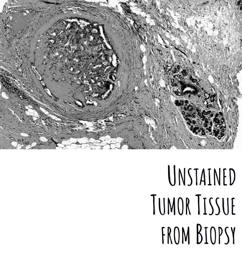
THE PROBLEM.
Cancer diagnosis is traditionally done on intraoperative frozen tissue sections by post-surgical histopathologic analysis and, in selected cases, by elaborated and time-consuming molecular diagnosis. The analysis of the biopsy is performed through the staining of the tissue and the evaluation of the morphology of its cells under an optical microscope. This approach is neither fast nor quantitative, has an intrinsic variability in the interpretation depending on the experience of the histopathologist, and provides limited molecular information.
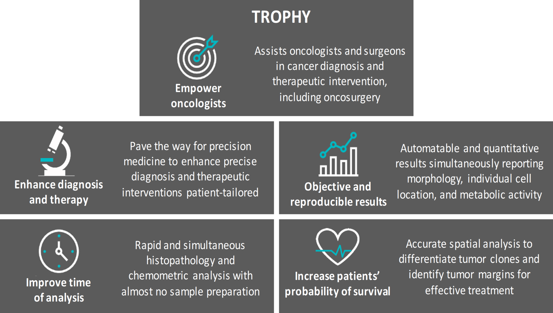
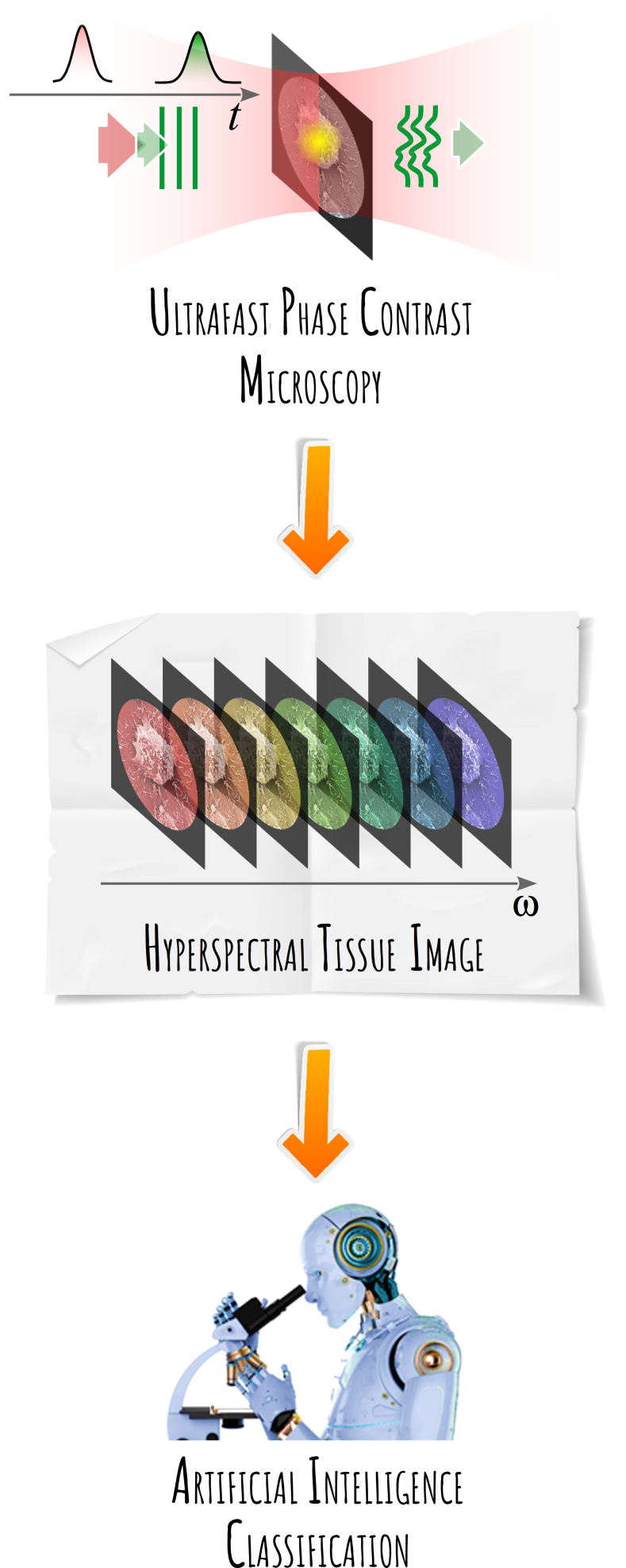
THE SOLUTION.
The EU-funded TROPHY project aims to develop a novel label-free vibrational microscopy approach that can image molecular biomarkers with unprecedented speed and chemical selectivity for a rapid, precise, and non-biased tumor analysis. To this purpose, the TROPHY microscope will blend in a unique fashion elements of several microscopies developed in the past decades, namely photo-thermal infrared, Fourier transform infrared and Digital Holography Microscopy, bringing them to the unprecedented ultrafast timescale. It will also integrate Artificial Intelligence to produce fast results and assist in the tumour grading process even during surgery.
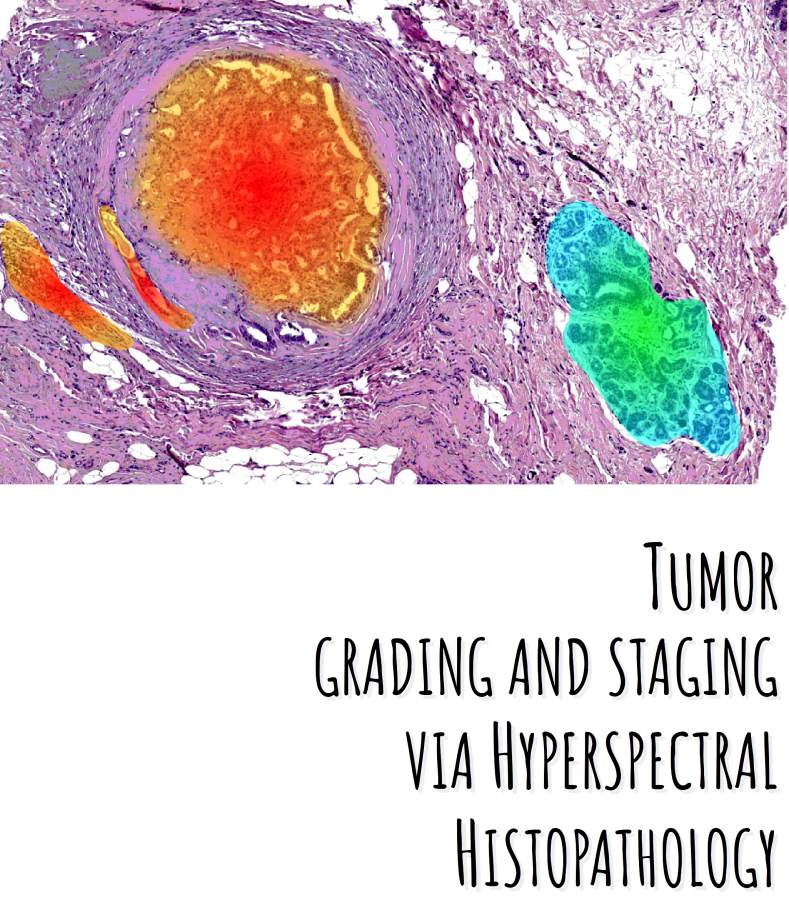
THE APPLICATION.
The TROPHY microscope will be used to assist healthcare professionals during tumor biopsy diagnostics, provide an accurate diagnosis for curative oncosurgery, guarantee complete resection during intervention, determine the best therapeutic approach tailored to the patient, and identify resistant tumor clones under targeted therapy, paving the way for continual precision medicine in cancer.
THE COMPETITIVE ADVANTAGE.
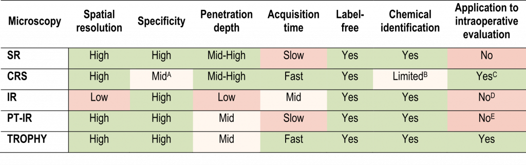
THE VISION.